Figure 2.
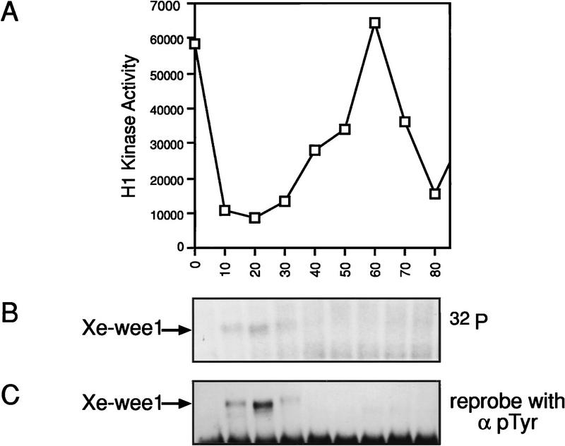
Xe-Wee1 has autophosphorylation activity in the first 30 min of the first mitotic cell cycle. The first mitotic cell cycle was initiated by treating unfertilized eggs with calcium ionophore (A23187). Ten eggs were collected at 10-min intervals and lysates were prepared in modified EB. Histone H1 kinase assays were used to monitor the progression through the cell cycle (A). Xe-Wee1 was immunoprecipitated (Ab 725:Ab 1532) from the remainder of each sample, and subjected to an immune complex autokinase assay with [γ-32P]ATP. The reactions were processed for SDS-PAGE, and the proteins were transferred to Immobilon membranes. After exposure to autoradiographic film (B), the filters were processed for immunoblotting with an anti-phosphotyrosine antibody (C).

