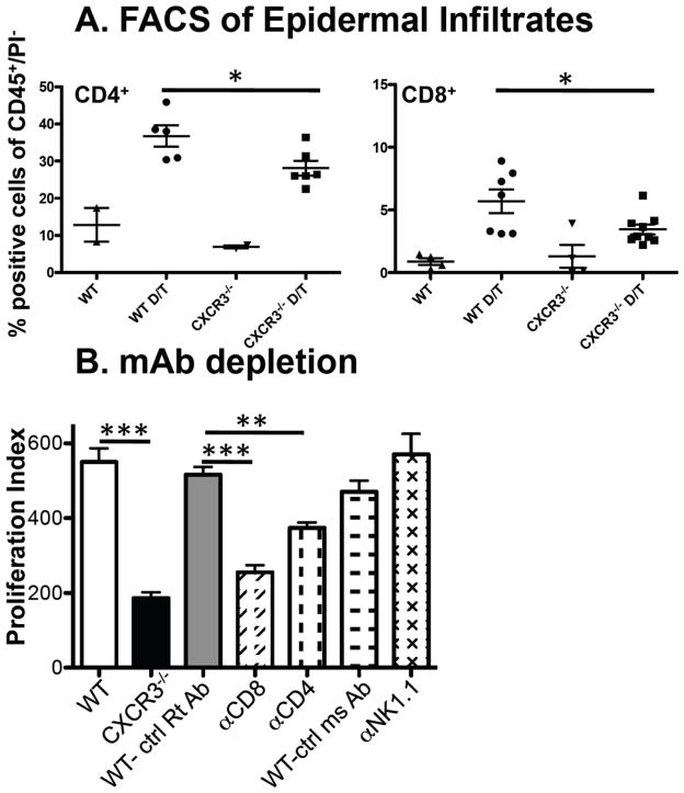
Figure 5.
T cell infiltration is reduced in CXCR3−/− compared to WT mice and T cell depletion in WT mice attenuates epidermal proliferation. (A) FACS analysis of DMBA/TPA treated skin reveals reduced CD4+ and CD8+ T cells in CXCR3−/− compared to WT mice. Epidermal preparations were generated from untreated WT and CXCR3−/− mice or short course DMBA/TPA treated skin (designated as D/T) and FACS analysis was performed for CD4+ and CD8+ T cells (data shown) and CD11b+ cells, Gr1+CD11b+cells and γδ/vγ5+ cells (data in Supplementary Figure 4). Each point represents an individual mouse and data are expressed as percentage of cells relative to total CD45+/PI− cells and revealed significant reductions in CD4+ and CD8+ T cells (*p<0.05). (B) WT C57BL/6 mice were treated with the indicated mAbs and DMBA/TPA treated skin was assessed for proliferation (***p<0.001 for WT (n=4) versus CXCR3−/− (n=3), ***p<0.001 for control IgG (n=2) versus anti-CD8 (n=7) and **p<0.01 for control IgG (n=2) versus anti-CD4 (n=6), combined data from 3 experiments).

