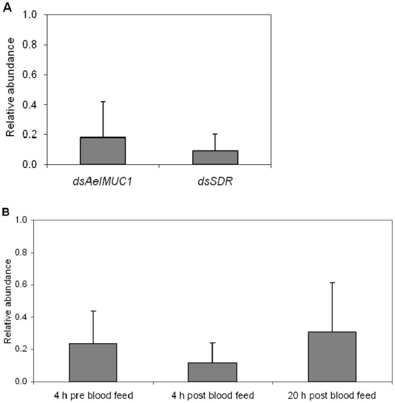
Figure 1.

Relative abundance in transcript levels of AeIMUC1 and SDR genes in female midguts of Aedes aegypti injected with the corresponding dsRNAs compared with control mosquitoes injected with dsβ-gal RNA. Transcript data for control and test genes were normalized using RpS17. (A) Results represent means ± SD for real-time PCR analysis of four replicates in each assay. Quantifications were performed at 24 hours after blood feeding. (B) Results represent means ± SD for AeIMUC1 gene real-time PCR analysis of three replicates. Quantifications were performed at 4 hours before and 4 hours and 20 hours after blood feeding.
