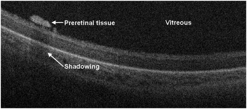
Figure 2.

Cross-sectional SD OCT image shows a laterally elongated preretinal structure with shadowing. As these preretinal structures were sometimes found adjacent to blood vessels, we hypothesize that they may represent early proliferative changes. In the sessions where preretinal tissue was seen on SDOCT (presumably within zone I) the ROP was staged as 3 or greater in 38% of sessions.
