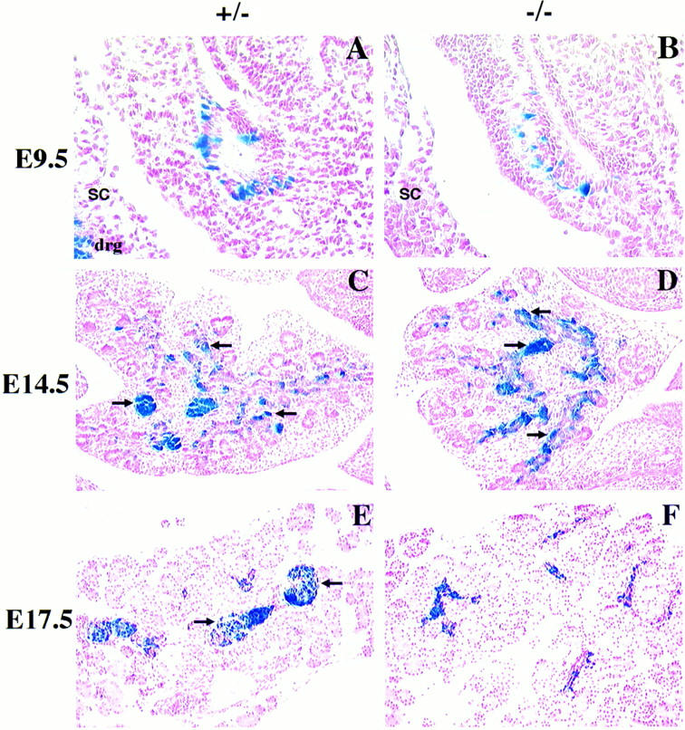
Figure 2.

Abnormal pancreatic islet morphogenesis in BETA2 −/− mice. (A–D) Sagittal sections of BETA2 +/− (left panels) and BETA2 −/− (right panels) embryos at E9.5 (A,B) and pancreas at E14.5 (C,D) stained with X-gal. There were no obvious differences in morphology of the early developing pancreas or in the number of β-gal-positive cells. β-Gal-positive cells were similarly distributed in both +/− and −/− tissue. β-Gal expression was also detected in both the dorsal and ventral pancreas. (E,F) Pancreata of +/− and −/− mice at E17.5 stained with X-gal. At E17.5 (E) there was evidence of islet formation in the +/− pancreas, as indicated by β-gal-positive cells, which formed a sphere-shaped structure with a distinct border surrounded by acinar cells (arrows). In contrast, distinct islets were not present in −/− pancreas at E17.5 (F), although β-gal-positive cells were seen without distinctive shape or border. Sections were counterstained in nuclear fast red. (drg) Dorsal root ganglion; (sc) spinal cord. Original magnification, 500× (A,B); 250× (C–F).
