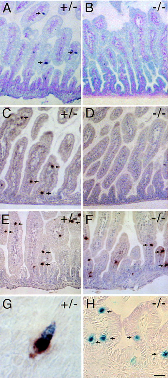
Figure 8.

Absence of enteroendocrine cells expressing secretin and cholecystokinin in BETA2 −/− mice. Immunostaining for secretin (A,B), CCK (C,D), and serotonin (E,F) (arrows) in the small intestine of 2 day old BETA2 +/− (left panels) and BETA2 −/− (right panels) mice. Note the absence of secretin- and CCK-staining cells in the −/− mice (B,D) compared to +/− mice (A,C). Nuclear β-galactosidase staining in +/− (G) and −/− (H). G shows a single cell with brown cytoplasmic staining for secretin and blue nuclear X-gal staining from an adult BETA2 +/− mouse. Several isolated mucosal epithelial cells (arrows) in the small intestine of a −/− mouse showing nuclear β-galactosidase activity. Bar, 100 μm for A–D; 50 μm for E,F; 5 μm for G; and 20 μm for H. Sections were stained using the ABC method with True Blue peroxidase substrate counterstained with contrast red (A,B) or DAB substrate counterstained with hematoxylin (C–H).
