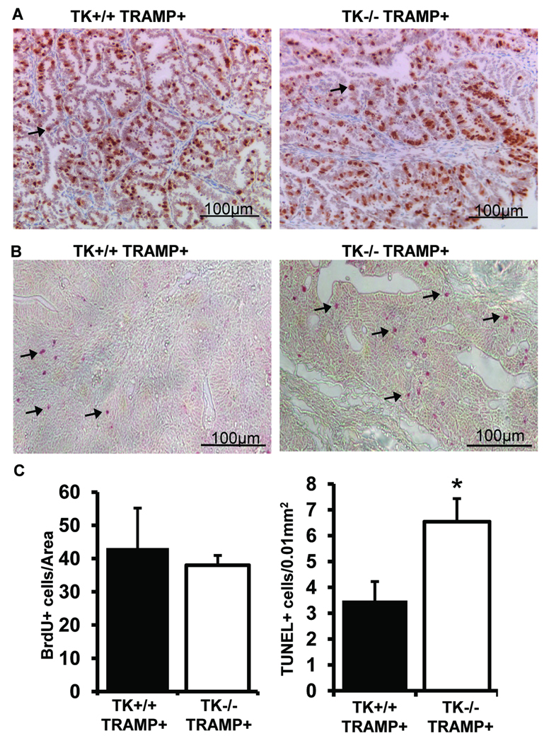
Figure 4. Prostate cell proliferation and apoptosis in TK+/+ TRAMP+ and TK−/− TRAMP+ mice.
A, BrdU immunostaining was performed on prostates from 30-week TK+/+ TRAMP+ and TK−/− TRAMP+ mice. B, Detection of TUNEL positive cells in prostate tissue of TK+/+ TRAMP+ and TK−/− TRAMP+ mice at 30-weeks of age. C, Prostates from 30-week-old TK+/+ TRAMP+ and TK−/− TRAMP+ mice did not exhibit differences in BrdU staining but did contain significant differences in the extent of TUNEL positive cell staining. Data are expressed as means ± SE. Five separate areas were counted from four independent specimens per group, and representative images are shown. *p<0.05 compared to TK+/+ TRAMP+ group. Arrows depict a few of the positive staining cells.

