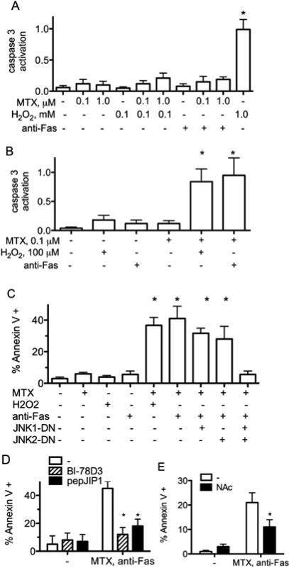
Figure 1.
MTX ‘primes’ Jurkat cells for increased sensitivity to apoptosis. Jurkat cells,A, were cultured for 48 hr. with MTX, H2O2, or 50 ng/ml anti-Fas and levels of caspase 3 activity determined. B, were cultured for 48 hr. with the indicated concentrations of MTX, followed by culture with H2O2 or anti-Fas for 24 hours before determination of caspase 3 activities. C,JNK1-DN, JNK2-DN, or ‘empty vector’ plasmids with a GFP plasmid were introduced by transient transfection. Cells were cultured for 48 hr. with or without MTX, 10-7M, and treated with anti-Fas for an additional 6 hr. Percent Annexin V positive cells were determined by flow cytometry after gating on GFP-positive and GFP-negative cells ± S.D. D, were cultured for 48 hr. with MTX, 10-7 M, in the presence or absence of BI-78D3 or pepJIP1, followed by culture with anti-Fas for 6 hours.Percent Annexin V positive cells were determined by flow cytometry ± S.D. E, were cultured for 48 hr. with MTX, 10-7 M, in the presence or absence NAc, followed by culture with anti-Fas for 6 hrs.Percent Annexin V positive cells were determined by flow cytometry ± S.D.* P < 0.05 relative to protein levels in untreated Jurkat cells.

