
Figure 2.
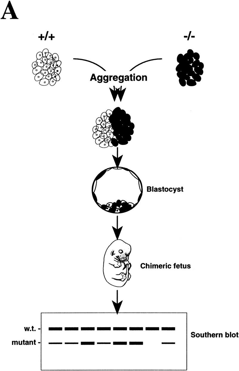
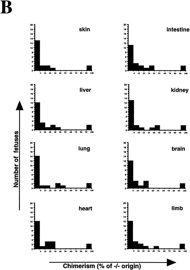
Proliferation of PARP mutant cells in vivo. (A) The diagram shows the experimental approach to examine the proliferation capacity and competence of PARP mutant cells. Wild-type (w.t., +/+) and PARP mutant (−/−) embryos were aggregated at the morula stage. The proliferation status of mutant cells in chimeric embryos was monitored by the contribution of each origin, which was determined by quantitative Southern blot analysis using DNA isolated from different organs (see also Materials and Methods). (B) A total of 21 E17.5 fetuses was obtained and eight tissues from each were analyzed by Southern blotting. The contribution from PARP−/− origin is represented by the percentage along with the number of fetuses that contained the same range of contribution in a given organ.