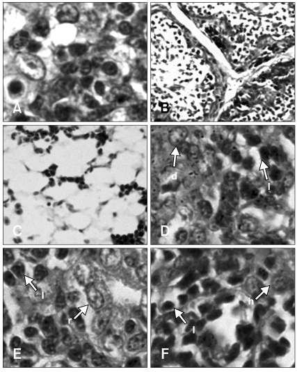
Fig. 2.
Histopathology lesions from the ALV-J infected group at 4-weeks-of-age. No difference was found between group 1 (day 1 infection) and group 2 (day 7 infection). Depletion of lymphocytes in the thymus (A) and bursa of Fabricius (B). All types of cells in the bone marrow were depleted (C). Proliferation of dendritic cells in the spleen (D). Lymphocytes infiltration in the kidney (E) and liver (F). H&E stain. A, D, E and F: ×1,000, B: ×100, C: ×200. d: dendritic cell, h: hepatocyte, l: lymphocyte, r: renal tubule.

