
Figure 6.
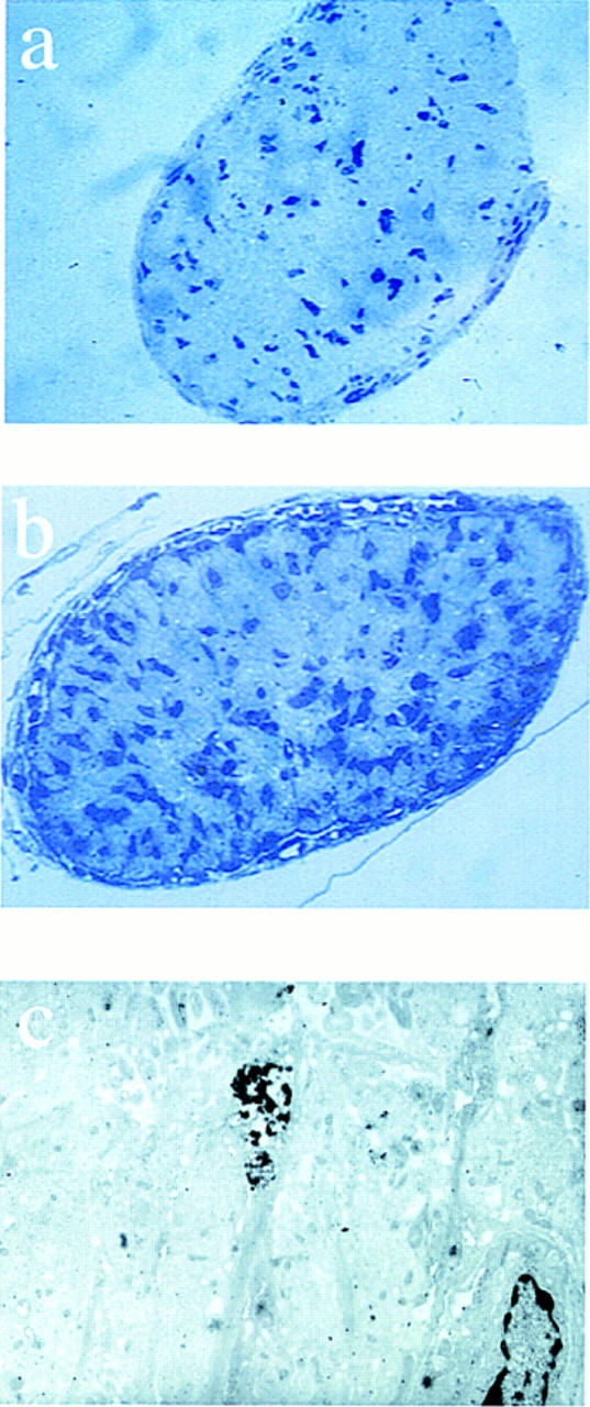
Glial cell number is increased in the optic nerve. Semithin sections (a,b) and electron microscopy of optic nerve from 6-day-old neonatal mice (c). Sections were cut 1 mm anterior of the optic chiasm and stained with toluidine blue and the number of nuclei were counted. Representative sections are shown for p27+/+ (a) and p27−/− mice (b). Electron microscopy of ultrathin sections from p27−/− mice revealed the presence of several apoptotic nuclei. A representative field (c) showing two nuclei (lower right and center of the field), one of which is undergoing apoptosis (center of the field).