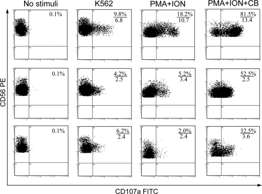
Fig. 1.
Typical pattern of the degranulation of NK cells from a healthy donor (upper row) and a patient with FRH in recurrence (middle row) and in remission (lower row) without stimulation and in response to K562 cells, phorbol 12-myristate 13-acetate plus ionomycin (PMA+ION), and phorbol 12-myristate 13-acetate plus ionomycin plus cytochalasin B (PMA+ION+CB). NK cell degranulation assay was performed as described in Materials and Methods, and the results were assessed by flow cytometry. Shown are the results for gated CD3− CD56+ NK cells. The numerators indicate percentages of NK cells that have externalized CD107a during the 4-h incubation period, and the denominators indicate the mean fluorescence intensity (MFI) of CD107a on CD107a+ NK cells (in arbitrary units).

