
Figure 4.
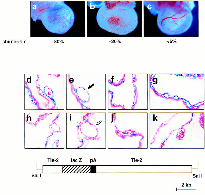
SCL−/− cells contribute to the endothelium of small but not large vessels in chimeric yolk sacs. The contribution of SCL-deficient cells to the endothelium of E9.5 chimeras was analyzed by β-galactosidase whole-mount staining. (a–c) β-Galactosidase-stained yolk sacs derived from SCL−/−/ROSA chimeras. The approximate degree of chimerism is shown below each. Note the disorganized vascular architecture in the higher percentage chimeras, a and b. Original magnification, 22.5×. (f–k) Sections of stained yolk sacs of chimeric embryos derived from Tie-2–lacZ-marked SCL+/− or SCL−/− ES cells. (d,e) SCL+/− cells contribute uniformly to capillaries and vitelline vessels in chimeric yolk sacs. Note the contribution of lacZ-positive cells to large vessels, as indicated by the solid arrow in e. (f–k) In contrast, SCL−/−-derived endothelial cells were not detectable lining the lumens of vitelline vessels, as highlighted by the open arrow in i. Note that the SCL−/− ES cells contribute efficiently to capillaries. Original magnification, 400×, except for a and f, which were 200×. The structure of the Tie2–lacZ transgene (kindly provided by T. Sato, University of Texas Southwestern Medical School, Dallas) is shown in the bottom panel.