
Figure 4.
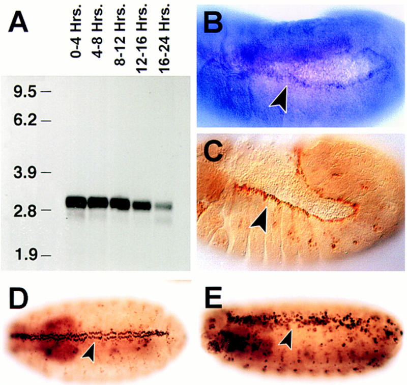
puc expression: Its modulation by puc activity. (A) Northern analysis of puc RNA expression in embryos at various times during development. (A) 0–4 hr; (B) 4–8 hr; (C) 8–12 hr; (D) 12–16 hr; and (E) 16–20 hr. The 2.9-kb puc transcript is apparent. (B) puc RNA detected in stage 13 by whole mount in situ hybridization. The expression in the dorsal-most epidermal cells is indicated. (C) stage 13 pucE69 heterozygous embryos stained with an antibody against β-galactosidase. The arrowhead points to the cells of the leading edge of the epidermis expressing β-gal. Notice that these are the same cells as in B. At early stages, evident puc expression is present in amnioserosa cells. (D) β-galactosidase expression of puc320 heterozygous embryos (stage 14). (F) A considerably higher number of cells, and at higher levels, express β-galactosidase in puc320/puc320 stage 15 embryos (arrowhead).