
Figure 6.
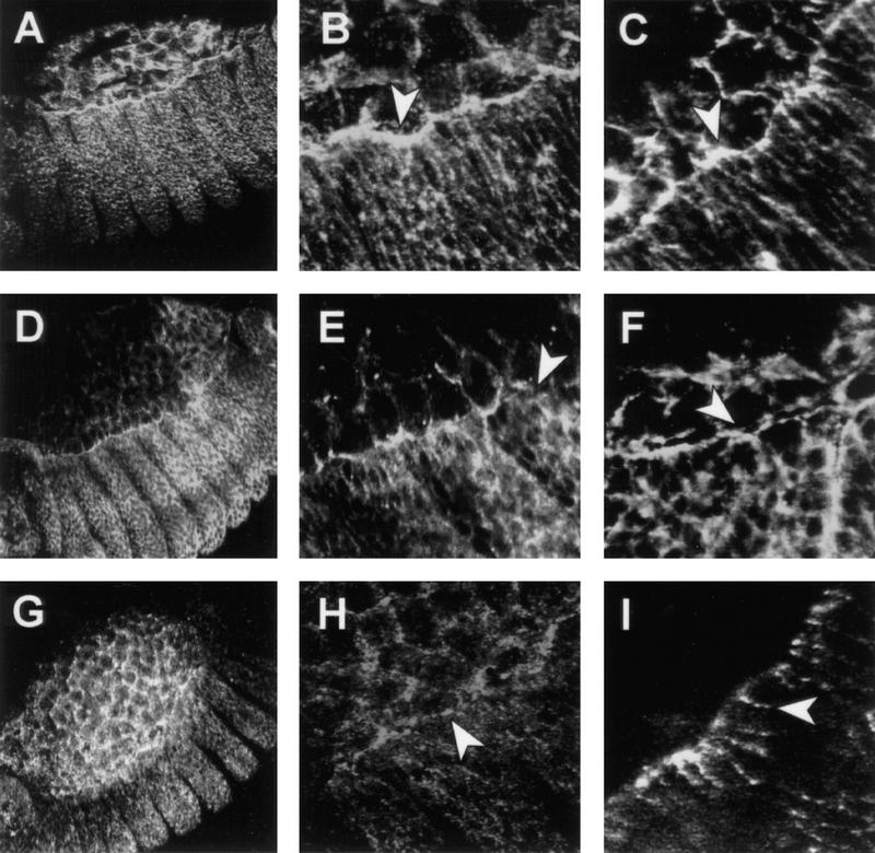
puc controls the accumulation of actin and myosin in the leading edge of the epidermis during dorsal closure. Confocal fluorescent micrographs of the boundary between the amnioserosa and the epidermis in stage 13 embryos. The distribution of nonmuscle myosin (A,B,D,E,G,H) and filamentous actin (C,F,I) are shown in wild-type embryos (A–C), pucE69 embryos (D–F) and ArmGal4/UASpuc embryos (G–I). Embryos were stained for filamentous actin with phalloidin or for nonmuscle myosin with antibodies. Whereas actin and NMM are accumulated along the leading edge in wild-type embryos (arrowheads in B and C), in puc mutants their level decreases and it is possible to observe gaps (arrowheads in E and F) between the amnioserosa and the epidermis. After Puc overexpression, NMM is maintained in the amnioserosa (arrowhead in H) but the level of expression in the epidermis is severely reduced. Actin ceases to be expressed in the amnioserosa and it appears on patchy spots in the epidermis (arrowhead in I).