
Figure 7.
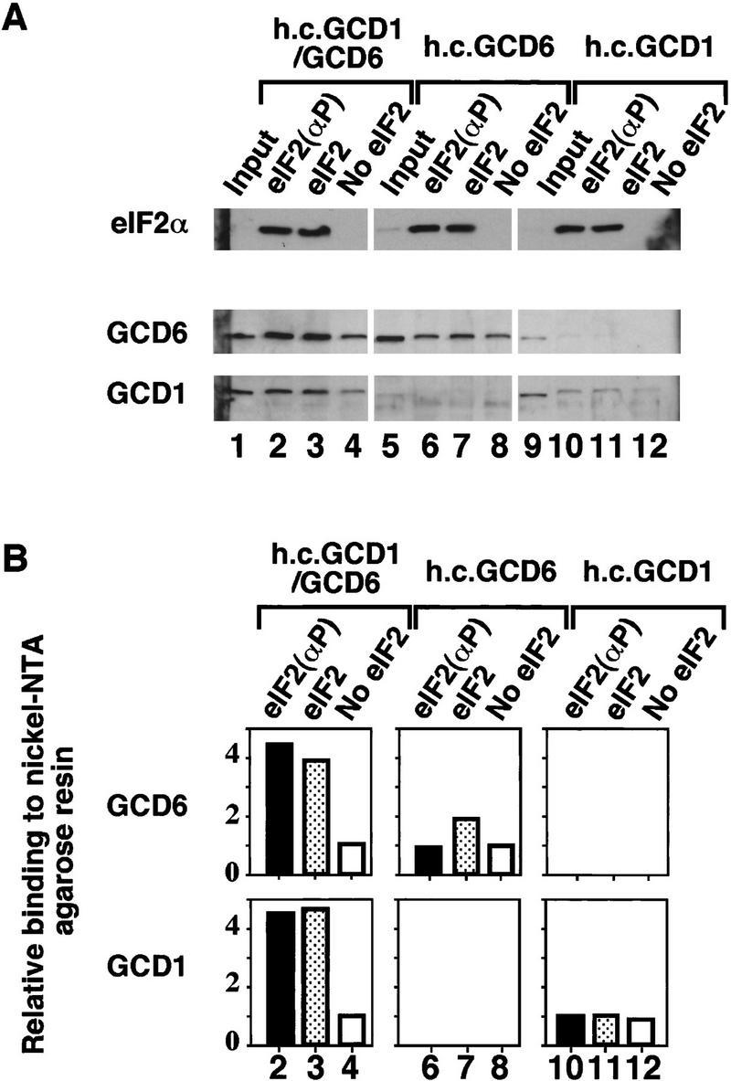
Binding of the catalytic GCD1/GCD6 subcomplex to His-tagged eIF2. As described for Fig. 4 except that the cell extracts used contained the overexpressed catalytic GCD6/GCD1 subcomplex (lanes 1–4) or overexpressed single subunits GCD6 (lanes 5–8) or GCD1 (lanes 9–12). (A) Western blot analysis of eIF2α and eIF2B subunits. The binding of 33 μg of cell extracts to Ni-NTA–agarose beads in the presence of purified His-tagged eIF2 prephosphorylated with HCR kinase (lanes 2,6,10), unphosphorylated purified His-tagged eIF2 (lanes 3,7,11), or without added purified His-tagged eIF2 (lanes 4,8,12). (Input) 10 μg of each cell extract used (lanes 1,5,9). (B) Histograms showing densitometry of signals for each eIF2B antibody shown in pellet lanes (lanes 2–4,6–8,10–12,14–16) from A relative to the density of the signal in lanes 4, 8, and 12, which were assigned an arbitrary value of 1.