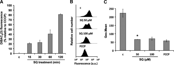
Fig. 2.
Effects of SQ on ΔΨp and ΔΨm. (A) Promastigotes were incubated without (c) and with 100 μM SQ in HBS for 15, 30, 60, and 120 min at 28°C and then treated with a 1 μM concentration of the specific plasma membrane potential probe DiBAC4(3) for 10 min at 28°C. DiBAC4(3) fluorescence is represented relative to that of parasites treated with 10 μM CCCP, used as 100% depolarization of the plasma membrane potential. Results are means ± standard deviations (SD) for three independent experiments. (B and C) SQ-induced ΔΨm depolarization. L. donovani promastigotes were treated without (c) and with 50 and 100 μM SQ for 15 min, stained with 0.8 μM Rh123, and analyzed for fluorescence by flow cytometry. Parasites treated with 10 μM FCCP for 10 min were used as a depolarization control. (B) Histograms from a representative experiment of three independent experiments. (C) Geometric mean (Geo.Mean) channel fluorescence values ± SD for three experiments. The experimental values were significantly different from control values by Student's t test (P < 0.02). *, geometric mean for the 47% of parasites that showed an Rh123 accumulation decrease compared to the control.

