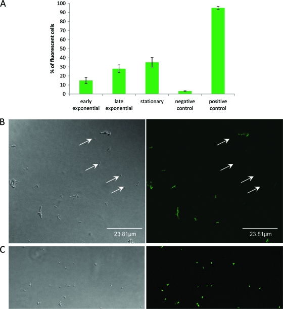
Fig. 1.
Individual cell measurement of Agr expression during growth of L. monocytogenes ARD009 in homogenized liquid cultures incubated at 25°C. (A) Percentages of GFP fluorescent cells detected by flow cytometry. A positive GFP signal is detected in Agr-ON cells (cells expressing Pagr-gfp). (B) Phase-contrast microscopy and fluorescence microscopy of L. monocytogenes ARD009 cells expressing Pagr-gfp after 16 h of incubation at 25°C. (C) Phase-contrast microscopy and fluorescence microscopy of L. monocytogenes EGD-e(pNF8-GFP). White arrows show examples of Agr-OFF cells.

