Fig. 3.
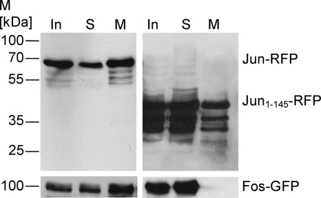
Immunoblots of immunoprecipitation fractions of BHK cell extracts. Immunoblot analysis of Jun-RFP probed with an anti-RFP antibody show signals in all three fractions of immunoprecipitation (top left). Coprecipitated Fos-GFP is also detectable in each fraction due to the interaction of Jun and Fos (bottom left). Immunoblot of truncated Jun1-145-RFP shows signals in each fraction of the immunoprecipitation (top right), whereas Fos-GFP is not detectable in the magnetosome fraction due to the loss of the Fos binding site in truncated Jun1-145 (bottom right). M, molecular mass.

