Figure 1.

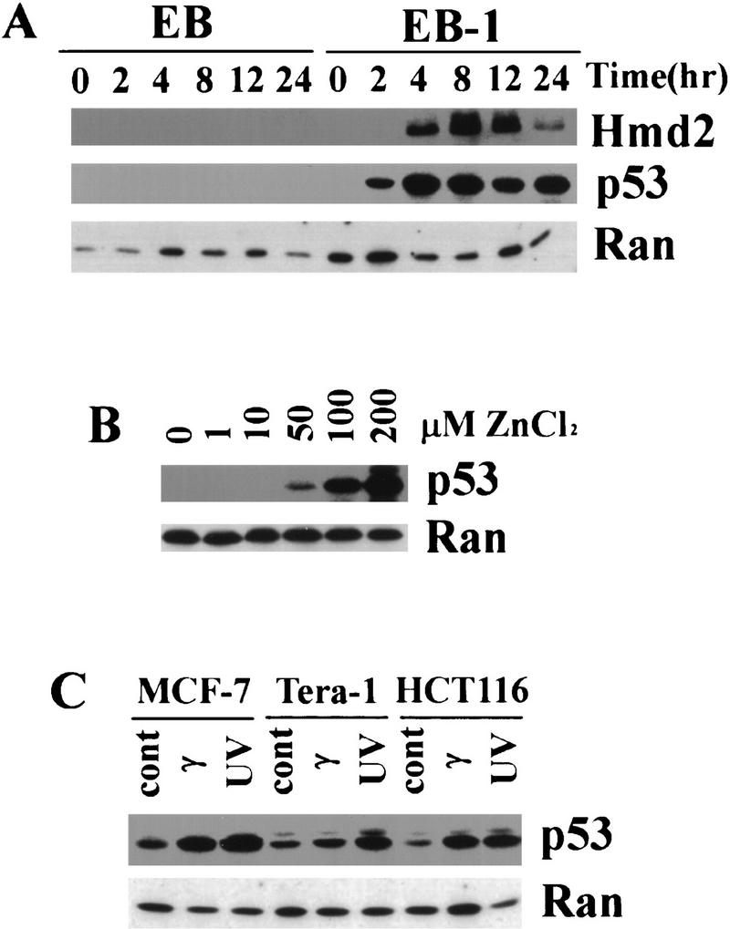
Inducible p53 in colon carcimona cell line EB-1. p53 and human MDM2 were detected by Western blots using monoclonal antibodies 1801 and 2A9, respectively. A small GTPase Ran was used as control. (A) EB-1 cells were stimulated with 100 μm zinc chloride starting at time 0 hr, and the level of p53 protein was elevated as early as 2 hr. Maximum induction of p53 was seen between 4 and 8 hr. Human MDM2 protein was also induced in these cells with a delay compared with p53 induction. No induction of p53 or hMDM2 in parental EB cells was observed following the same stimulation. The amount of 10 μg of total protein was loaded in each lane, and the same blot was probed by anti-p53, anti-hMDM2, and anti-Ran antibodies. (B) The levels of induced p53 protein in EB-1 cells depend on the zinc concentration in the culture media. EB-1 cells were cultured in presence of 0, 1, 10, 50, 100, and 200 μm of zinc chloride for 8 hr. A minimum of 50 μm of zinc was required to induce p53 proteins in these cells, and 100 and 200 μm zinc induced p53 to much higher levels in a dose-dependent manner. The amount of 20 μg of total protein was loaded in each lane. (C) Induction of p53 by UV and γ irradiation in different cancer-derived cell lines. Each cell line was treated with radiation as indicated, and cell lysates were prepared after 3–6 hr (see Materials and Methods). p53 was induced following both UV and γ irradiation in all the cell lines tested. The amount of 65 μg of total protein was loaded in each lane.
