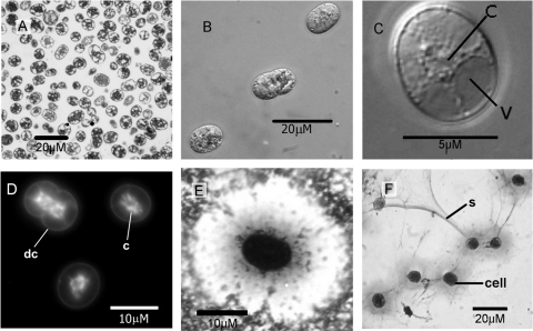
Fig. 4.
Cell morphologies of cells collected in February 2010 (salinity, >200). (A) Thin section of resin-embedded cells, visualized by low-magnification TEM. (B) Live cells viewed by Nomarski differential interference contrast with Z-stacking of several levels of focus, displaying the prominent granular nature of the cytoplasm. (C) Phase-contrast micrograph with Z-stacking of a single cell, showing typical peripheral vacuoles (v) with granular cytoplasm (c) constrained to a star-shaped mass at the center of the cell. (D) DAPI-stained cells viewed by epifluorescence microscopy, showing cytoplasm (c), distinguished by reticulate and rod-shaped, brightly fluorescent DNA. dc, dividing cell. (E) Typical capsular structure, visualized by negative staining (skim milk background) by a modification of the Anthony method (3). The central, deeply stained cell is surrounded by a lightly stained capsule against the dark background of stained milk proteins. (F) Demonstration of slime production using the Anthony method (3) for direct staining of slime and capsules. Capsules are visible around the deeply stained cells as fainter areas of staining, with prominent slime strands (s) extending between cell groups.

