
Figure 4.
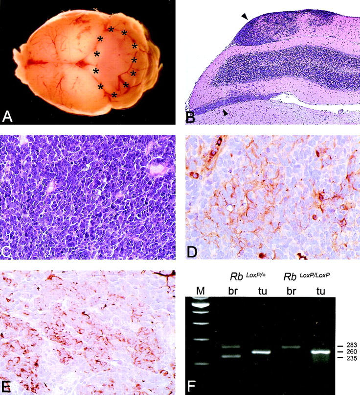
Histopathological analysis of medulloblastomas in GFAP–Cre;RbLoxP/LoxP;p53−/− mice. (A) Macroscopical appearance of a medulloblastoma arising in the cerebellar vermis of a 16-week-old mouse. (B) Low-power magnification of a tumor diffusely infiltrating in a cerebellar folia and in the subarachnoidal space (upper and lower arrows, respectively; H&E). (C) High-power magnification of a hypercellular tumor consisting of densely packed, polygonal cells with scant cytoplasm and hyperchromatic nuclei (H&E). Areas of early neuronal (MAP-2; D) and glial (GFAP; E) differentiation are detectable. (F) PCR analysis of Rb recombination in tumors arising in GFAP–Cre;RbLoxP/+;p53−/− and GFAP–Cre;RbLoxP/LoxP;p53−/− mice. The 283-, 260-, and 235-bp products correspond to the floxed (nonrecombined), the recombined, and the wild-type allele, respectively. The primers used for the analysis (Rb212, Rb19E, and Rb18) are the same as described in Fig. 3.