
Fig. 2.
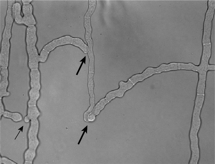
Photograph of the wild-type strain showing fusion hyphae. The wild-type strain was grown between two sheets of cellophane, and a region behind the growing edge of the colony containing fusion hyphae was photographed. The thicker arrows show locations where the fusion hyphae have completed the process of cell fusion. The thinner arrow points to two fusion hyphae that are growing toward each other but have not yet fused.