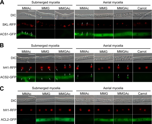
Fig. 7.
Cellular localization of ACS1-GFP (A), ACS2-GFP (B), and ACL2-GFP (C) depending on the culture conditions. Submerged mycelia were harvested for microscopic observation 24 h after conidium inoculation in liquid minimal medium supplemented with potassium acetate (MMAc), glucose (MMG), and both potassium acetate and glucose (MMGAc). Aerial mycelia were collected 3 days after inoculation on solid MMAc, MMG, MMGAc, and carrot agar (Carrot). The middle panels show RFP-SKL and hH1-RFP localized in peroxisomes and nuclei, respectively. All of the white arrows indicate peroxisomal localization (A) and nuclear localization (B and C). DIC, differential interference contrast. Scale bar, 20 μm.

