
Fig. 4.
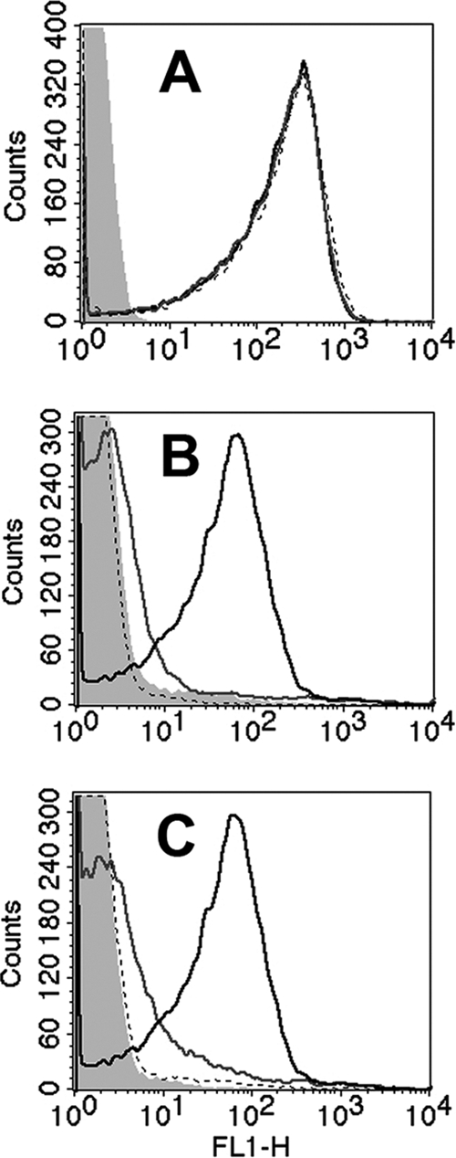
Binding of fH to fHbp and NspA KO mutants of group B strain H44/76 by flow cytometry. (A) Binding of anti-PorA MAb (P1.7, 20 μg/ml, positive control). Black line, wild-type strain; gray line, fHbp KO mutant; dashed line, double fHbp and NspA KO mutant; filled gray area, no MAb. The binding curves of the three MAbs are superimposed. (B) Binding of purified human fH (100 μg/ml). Designations are the same as in panel A except that the filled gray area is for no added fH. (C) Binding of fH in human serum (20%, IgG depleted). Designations are as in panel B. The results were replicated in two independent assays.