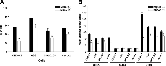
Fig. 12.
Cholesterol is important for CDT association and intoxication of cells. (A) Cells from the indicated lines were untreated or treated with 5 mM (for AGS cells) or 10 mM (for other cells) MβCD for 1 h at 37°C, followed by exposure to 200 nM CDT holotoxin for 48 h. Cell cycle distribution was analyzed using flow cytometry. (B) Cells from the indicated lines were untreated or treated with 5 mM (AGS cells) or 10 mM (other cells) MβCD for 1 h at 37°C, followed by incubation with the individual CDT proteins for 2 h at 4°C. The binding activity of each CDT protein was assessed by flow cytometry for FITC fluorescence. The results represent the means and standard deviations from three independent experiments. An asterisk indicates P < 0.05 compared to each the untreated MβCD group, as determined by Student's t test.

