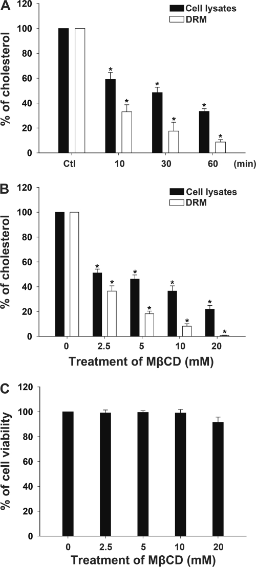
Fig. 4.
Cholesterol depletion in CHO-K1 cells by treatment with MβCD. (A) CHO-K1 cells were treated with 10 mM MβCD at 37°C and incubated for the indicated times. The cells were harvested and subjected to cold detergent extraction using 1% Triton X-100, followed by centrifugation to isolate the DRM fraction. The prepared total cell lysates and DRM fraction then were analyzed for cholesterol concentration as described in Materials and Methods. (B) CHO-K1 cells were treated with various concentrations of MβCD (0, 2.5, 5, 10, and 20 mM) for 1 h. Whole-cell lysates and the DRM fraction then were prepared for cholesterol level analysis. (C) Cell viability was barely influenced after treatment with 0 to 20 mM MβCD, as determined by the trypan blue exclusion assay. The data represent the means and standard deviations from three independent experiments. An asterisk indicates P < 0.05 compared to results for each untreated control group, as determined by Student's t test. DRM, detergent-resistant membrane; MβCD, methyl-β-cyclodextrin.

