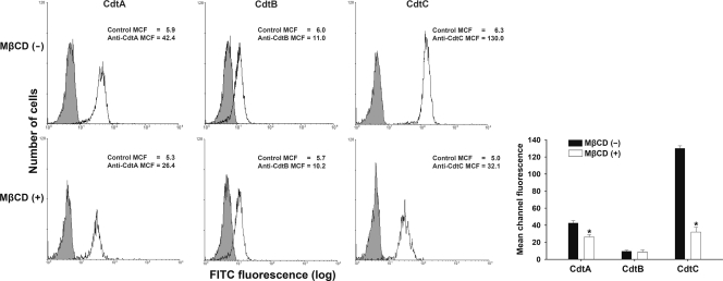
Fig. 5.
Sufficient cellular cholesterol is essential for CdtA and CdtC binding to CHO-K1 cells. The cells were left untreated (upper) or treated with 10 mM MβCD (lower) for 1 h at 37°C, followed by exposure to 200 nM the individual recombinant C. jejuni CDT proteins. After incubation with the individual CDT proteins for 2 h at 4°C, the cells were stained with control preimmune serum or individual antiserum against each CDT subunit and stained with FITC-conjugated anti-mouse IgG. Binding activity was assessed by flow cytometry. The numbers represent the mean channel fluorescence (MCF). The quantitative data represent the means and standard deviations from three independent experiments and are shown in the lower right panel. Statistical analysis was calculated using Student's t test compared to each untreated MβCD group. An asterisk indicates statistical significance (P < 0.05).

