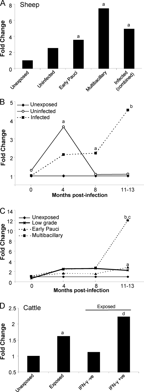
Fig. 5.
IDO gene expression in peripheral blood cells is increased with M. avium subsp. paratuberculosis infection and modulated throughout the disease course. qPCR results of IDO gene expression, normalized against the appropriate reference gene, in peripheral blood cells from sheep and cattle. Fold change is relative to gene expression in an unexposed (control) group. (A) Expression of IDO in PBMCs from naturally and experimentally infected sheep at later stages of disease (1.5 to 4 years of age). Unexposed sheep (control), n = 11; uninfected sheep (sheep exposed to M. avium subsp. paratuberculosis but uninfected at necropsy), n = 5; sheep with early paucibacillary lesions, n = 7; sheep with multibacillary lesions, n = 7; infected sheep (combined early pauci- and multibacillary), n = 14. (B and C) Time course of IDO gene expression in sheep from experimental-infection trial A. Unexposed sheep (control), n = 20; uninfected sheep, n = 10; infected sheep (combined early pauci- and multibacillary), n = 18; sheep with low-grade lesions (infected sheep with a lesion grade of 1 or 2), n = 5; sheep with early paucibacillary lesions, n = 8; sheep with multibacillary lesions, n = 10. (D) Expression of IDO in PBMCs from experimentally infected cattle (n = 20) and unexposed controls (n = 10). Exposed cattle were further subdivided into those that responded in an M. avium subsp. paratuberculosis-specific IFN-γ assay (IFN-γ + ve [where “+ ve” indicates “positive”]; n = 6) compared to those that did not respond (IFN-γ − ve; n = 14), performed at 5 months postexposure. For significant differences, “a” indicates a P of <0.05 in comparisons with unexposed controls, “b” indicates a P of <0.001 in comparisons with unexposed controls and an uninfected group, “c” indicates a P of <0.01 in comparisons with low-grade and early paucibacillary lesion groups, and “d” indicates a P of <0.001 in comparisons with unexposed and IFN-γ-negative groups.

