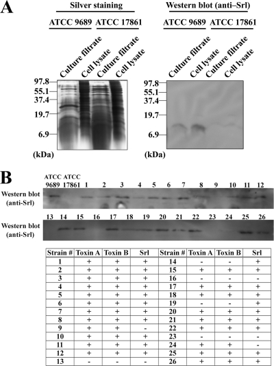
Fig. 3.
Production of Srl in C. difficile ATCC 9689 and clinical strains. (A) Whole-cell lysate and TCA-precipitated culture filtrate from C. difficile ATCC 9689 or C. novyi ATCC 17861 as the negative control were subjected to SDS-PAGE followed by silver staining. The same samples were analyzed by Western blotting with polyclonal anti-Srl antibody. (B) Whole-cell lysate of each strain was subjected to Western blotting with polyclonal anti-Srl antibody. For detection of toxins A and B, membrane-based enzyme immunoassays were performed. The results are summarized in the table; + and − denote positive and negative results, respectively, of enzyme immunoassay for toxins A and B or Western blotting for Srl.

