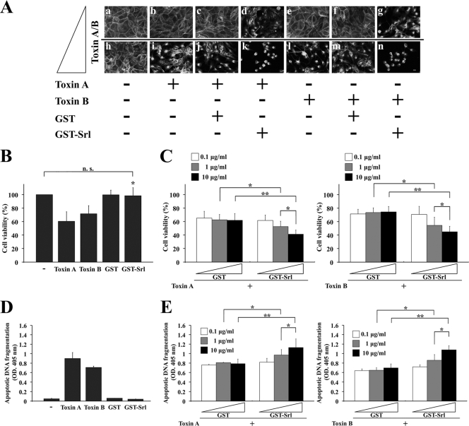
Fig. 5.
GST-Srl enhances the cytotoxicity of both toxin A and toxin B in epithelial cells. (A) Morphological changes of MDCK cells after treatment with toxin A (0.25 ng/ml [panels b, c, and d] versus 25 ng/ml [panels i, j, and k]) or toxin B (0.1 ng/ml [panels e, f, and g] versus 10 ng/ml [panels l, m, and n]) in the presence of either GST-Srl or GST. After 24 h of incubation, cells were fixed and labeled with TRITC-phalloidin. + and − denote positive and negative results, respectively. Bar, 10 μm. (B) MDCK cells were incubated with 250 ng/ml toxin A, 100 ng/ml toxin B, 10 μg/ml GST-Srl, or 10 μg/ml GST alone. After incubation for 24 h, cell viability was measured by an aqueous soluble tetrazolium/formazan assay and expressed as a percentage of the results from buffer-treated controls (−). All reactions were performed in triplicate, and the results are means ± SD of three independent measurements. n.s., not significant. *, P < 0.01 versus toxin A or toxin B. (C) MDCK cells were incubated for 24 h with the indicated concentrations of GST-Srl or GST in the presence of either 250 ng/ml toxin A or 100 ng/ml toxin B. The viability of MDCK cells was determined as in panel B. Results are means ± SD of three independent measurements. *, P < 0.05; **, P < 0.01. (D) MDCK cells were incubated with 250 ng/ml toxin A, 100 ng/ml toxin B, 10 μg/ml GST-Srl, or 10 μg/ml GST alone. After incubation for 24 h, lysates were prepared, and apoptotic oligosomal DNA fragmentation was assessed using a cell death ELISA kit. All reactions were performed in triplicate, and the results are expressed as optical density (OD.) at 405 nm. Mean values ± SD are indicated for three independent measurements. −, buffer-treated control. (E) MDCK cells were incubated for 24 h with the indicated concentrations of GST-Srl or GST in the presence of 250 ng/ml toxin A or 100 ng/ml toxin B. The cell death induced in MDCK cells was detected as in panel D. *, P < 0.05; **, P < 0.01.

