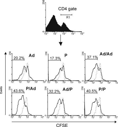
Fig. 4.
Proliferation assay performed on CFSE-labeled splenocytes of BALB/c mice immunized with P. vivax recombinant immunogens. The percentage of proliferation was calculated by flow cytometry on CD4+ gated splenocytes of mice immunized 2 weeks before with AMA-1 (continuous lines) administered as a recombinant adenoviral vector (Ad), as a protein in Montanide ISA720 (P), or as sequential inoculations of both immunogens in prime/boost protocols, compared to mice immunized with two doses of an irrelevant adenovirus control (dotted lines). CFSE-labeled splenocytes were induced to proliferate ex vivo for 30 h by adding PvAMA-1 to the cell culture. Plots are representative of two different experiments that included a total of six animals per group, analyzed in pools of three. Numbers represent mean values of the results.

