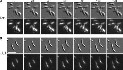
Fig. 4.
Time-lapse images of KR2616 (creS, creS::GFP+ JS14-xylX::ctpA) grown in PYEX with (A) or without (B) A22. DIC (top) and CreS-GFP fluorescence images (bottom) were taken at 20-min intervals. All cells were grown overnight in PYEX to induce excess CtpA. Cells in A were also treated with 50 μM A22 for 3 h before imaging. Scale bar, 5 μm. Outlines highlight a cell in which the crescentin structure was mobile.

