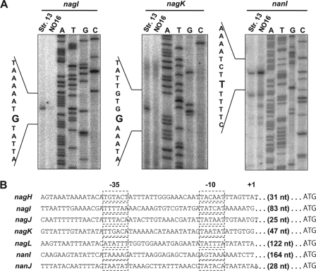
Fig. 8.
Determination of transcriptional start sites of nagI, nagK, and nanI. (A) Primer extension analysis of nagI, nagK, and nanIusing 32P-labeled primer and total RNA isolated from cultures of strains 13 and NO16 at the mid-exponential phase. The sequence around the transcriptional start site of each gene was determined by sequence reaction using the same primer. The transcriptional start sites are shown on the left. (B) Promoter sequences of hyaluronidase and sialidase genes in C. perfringensstrain 13 (1). The dashed boxes indicate −10 and −35 boxes. The ranscriptional start site is indicated by +1′. The length of the 5′ UTR of each gene is also indicated.

