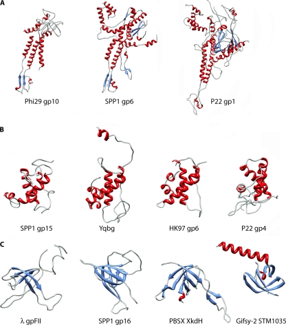
Fig. 3.
Conservation of the protein modules constituting bacteriophage connectors. (A) Gallery of portal protein structures determined by X-ray diffraction: ϕ29 gp10 (PDB accession no. 1FOU), SPP1 gp6 (PDB accession no. 2JES), and P22 gp1 (PDB accession no. 3LJ4). (B) Crystal structures of SPP1 gp15 (PDB accession no. 2KBZ), PBSX YqbG (PDB accession no. 1XN8), HK97 gp6 (PDB accession no. 3JVO), and P22 gp4 (PDB accession no. 3LJ4). (C) Crystal structures of λ gpFII (PDB accession no. 1K0H), SPP1 gp16 (PDB accession no. 2KCA), PBSX XkdH (PDB accession no. 3F3B), and Gifsy-2 STM1035 (PDB accession no. 2PP6). The coloring scheme used is based on secondary structures: blue, β-strands; red, α-helices; cyan, loops.

