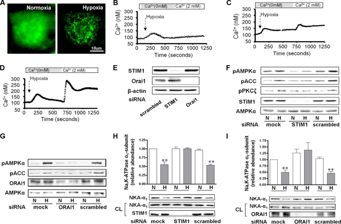
Fig. 3.
Hypoxia-induced STIM1 redistribution results in CRAC channel activity following AMPK activation and Na,K-ATPase downregulation. (A) A549 cells were transiently transfected with full-length WT STIM1-YFP and 24 h later were exposed 21% (normoxia) or 1.5% (hypoxia) O2, and the WT STIM1-YFP distribution was assessed. (B to D) Measurement of calcium in A549 cells transfected with siRNA against STIM1 (B), against Orai1 (C), or scrambled (D) and then exposed to hypoxia. Perfusion was started in Ca2+-free medium and switched to 2 mM Ca2+ as indicated. Results are from 4 experiments with 20 to 35 cells each. (E) Representative Western blot of the expression levels of STIM1 and Orai1 in A549 cells transfected with the respective siRNA. β-Actin was used as a loading control. (F and G) A549 cells were transfected with siRNA against STIM1 or Orai1 or with scrambled siRNA, and 48 h later cells were exposed to 21 or 1.5% O2 for 10 min. pAMPKα, AMPKα, pACC, pPKCζ, STIM1, and Orai1 protein levels are shown. (H and I) Cells transfected as for panel E were exposed to 21 or 1.5% O2 for 60 min, and the Na,K-ATPase α1 subunit plasma membrane (NKA-α1) abundance was determined. Representative Western blots of the NKA-α1 at the plasma membrane and in total cell lysates and STIM1 or Orai1 are shown. Results are means ± SEM (n = 4). **, P < 0.01.

