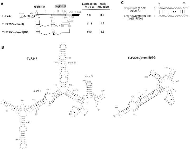
Figure 1.
Structure and expression of rpoH–lacZ gene fusion TLF247 and its derivatives. (A) Schematic diagram of TLF247 and its deletion derivatives. The locations of regions A and B are indicated by hatches. Segments of mRNA that correspond to each of the stem structures (I–IV; Fig. 1B) are shown above the diagram, and nucleotide numbers are shown below. Pulse-labeling (2 min) with [35S]methionine was performed before or 3 min after temperature upshift (30°C to 42°C), and immunoprecipitates were analyzed by SDS-PAGE as described in Materials and Methods. Synthesis rates were normalized to that of TLF247 labeled at 30°C. (Solid arrowheads) Positions of 2G substitutions. (B) mRNA secondary structures predicted for rpoH regions of TLF247 and TLF229Δ(stemIII)GG by mfold (Morita et al. 1999); the structures with minimum free energy are shown. The initiation codon, region A (nucleotides +6–20), SD sequence, and major stems (I–IV) are indicated. Region B (nucleotides +112–208) is shown as boldface letter. (●) G–U pairing. (C) Putative base-pairings between the downstream box (region A) of rpoH and the anti-downstream box of 16S rRNA (spanning 1469–1483 nucleotides).

