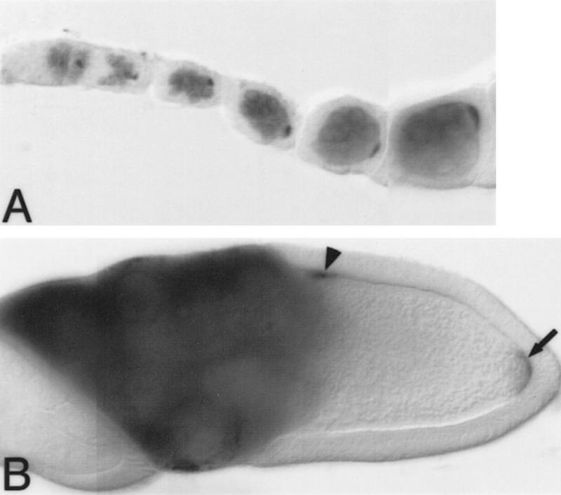
Figure 4.
Bru protein colocalizes with osk and grk mRNAs during oogenesis. Distribution of Bru protein in whole-mounted preparations of developing egg chambers. Each egg chamber contains a cluster of nurse cells on the left, and a single oocyte on the right, oriented with the posterior of the oocyte to the right. Bru protein is visualized as a dark stain. (A) Early oogenesis. Bru protein can be seen in all of the germ cells, beginning in stage 2A of the germarium, and is clearly accumulating preferentially in the presumptive oocyte by stage 2B. As with osk mRNA, Bru can be seen as a posterior crescent within the oocyte even in the early stages of oogenesis. Note that Bru protein is also maintained throughout the nurse cells, the site of osk mRNA synthesis, as oogenesis progresses. (B) Stage 10 egg chamber. As with osk mRNA (Ephrussi et al. 1991; Kim-Ha et al. 1991), the posterior localization of Bru in the oocyte becomes more striking in later egg chambers (arrow). Additionally, in stage 10 egg chambers Bru mimics the anterodorsal pattern of grk mRNA localization over the oocyte nucleus (arrowhead; Neuman-Silberberg and Schüpbach 1993).

