
Figure 5.
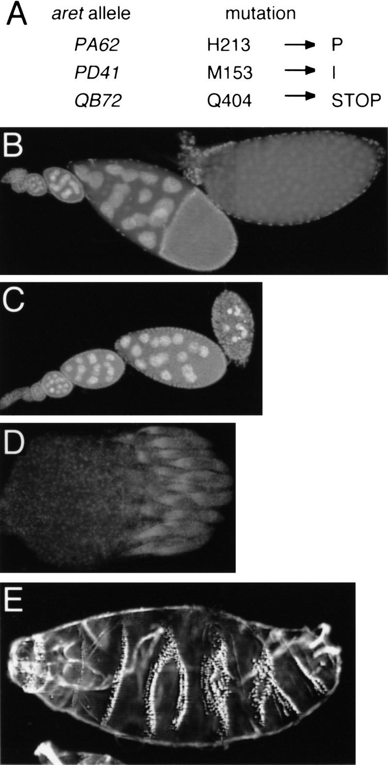
(A) Mutations in aret alleles. Amino acid positions refer to Fig. 3A. (B–D) Whole-mounted tissue stained with DAPI. (B) Wild-type ovariole composed of a string of progressively developing egg chambers, with the oldest egg chamber on the right. (C) Ovariole from an aretPA62/Df(2L)esc-P3-0 female. The progression of oogenesis appears normal until approximately stage 9, when the egg chambers deteriorate. No late-stage eggs are formed or laid. aretPD41/Df(2L)esc-P3-0 females have a similar phenotype. (D) A complete ovary, consisting of 15–20 ovarioles, from an aretQB72/Df(2L)esc-P3-0 female. All ovarioles are arrested in germarial stages. (E) Larval cuticle of an embryo from an aretPA62/aretPD41 mother showing complex defects in the pattern of segmentation (cf. Fig. 6A, wild-type cuticle). The severity of this phenotype is variable; the defects do not resemble the mutant phenotypes resulting from either over- or underexpression of osk.