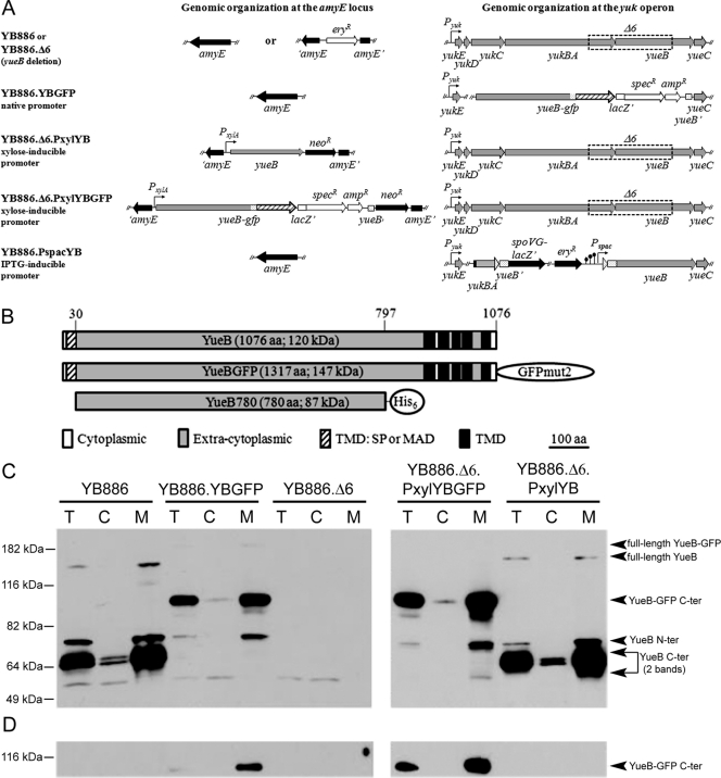
Fig. 1.
Expression of yueB and yueB-gfp fusions in B. subtilis. (A) Relevant genomic organization of strains used in the present study, with different levels of yueB or yueB-gfp expression (see Materials and Methods for details). The yueB-gfp fusion was cloned under the control of the native yukE promoter (strain YB886.YBGFP) or of the inducible PxylA promoter at the ectopic amyE locus (strain YB886.Δ6.PxylYBGFP). (B) Scheme of YueB, YueB-GFP, and YueB780 showing their putative signal peptide (SP) and transmembrane (TMD) segments, extracellular (ectodomain), and cytoplasmic regions. YueB780 is a dimeric elongated fiber active to trigger SPP1 DNA ejection in vitro whose sequence covers most of the ectodomain region of YueB (41). (C and D) Production of YueB and YueB-GFP in total cell extracts (T) and after fractionation into cytoplasmic (C) and membrane (M) fractions. Their composition was characterized by Western blotting with anti-YueB780 (C) and anti-GFP (D) antibodies. The amount of total protein loaded per lane was 30 μg (NanoDrop quantification) for YB886.Δ6.PxylYBGFP and YB886.Δ6.PxylYB (right panels), whereas 150 μg (5-fold) were loaded for YB886, YB886.YBGFP, and YB886.Δ6 (left panels) to increase sensitivity. Molecular mass markers are shown on the left. The assignment of YueB-derived bands is shown on the right.

