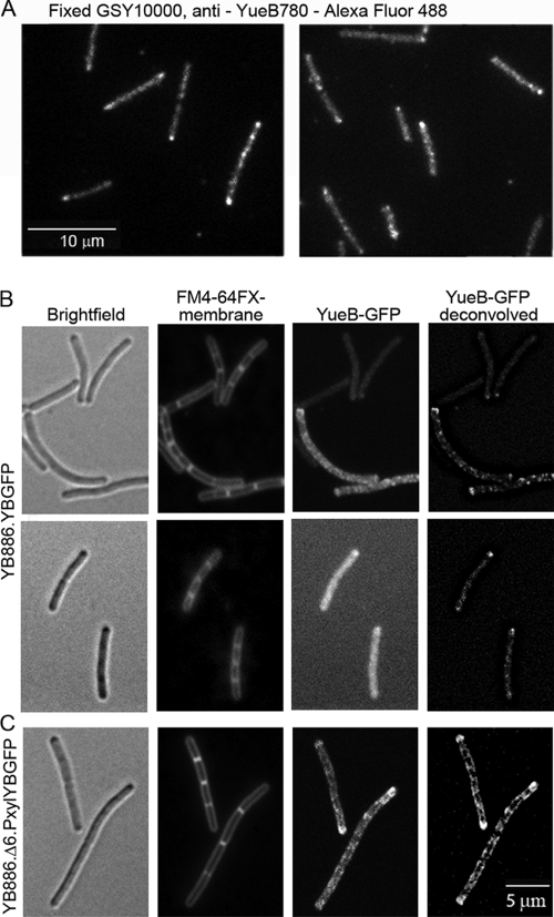
Fig. 4.
Localization of YueB in B. subtilis. (A) Labeling of YueB at the cell surface of fixed GSY10000 cells labeled with polyclonal anti-YueB780 antibodies. (B and C) Cytoplasmic localization of YueB in B. subtilis YB886.YBGFP (expression of the yueB-gfp fusion under the native Pyuk promoter; see Fig. 1A) (B) and YB886.Δ6.PxylYBGFP (yueB-gfp fusion under the control of a strong xylose-inducible promoter; see Fig. 1A) (C). The YueB cytoplasmic carboxyl terminus (39, 41) was fused to GFP (Fig. 1). The columns in panels B and C show, from left to right, bright-field images, FM4-64FX-stained membranes, YueB-GFP fluorescence, and two-dimensional deconvolution of the YueB-GFP signal.

