Fig. 6.
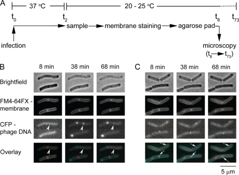
Time-lapse of bacteriophage SPP1delX110lacO64 infection. (A) Experimental setup: t0, infection point; t2, 2 min p.i., t8-73, 8 to 73 min p.i. (B and C) Images of B. subtilis producing LacI-CFP infected by phage SPP1delX110lacO64 (input multiplicity of 3) in the absence (B) or presence (C) of HPUra were taken at the postinfection times indicated. Rows: bright-field images, top row; FM4-64FX membrane staining, second row; LacI-CFP, third row. The bottom row shows overlays of membrane staining and CFP fluorescence. The arrowheads show a focus that disappears during infection. The white arrows show small LacI-CFP spots in infected cells treated with HPUra.

