
Figure 6.
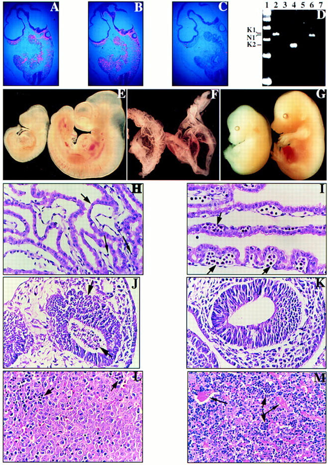
Phenotypic comparisons of N-ras−/−; K-ras+/− embryos and control littermates. In situ hybridization was performed on serial parasagittal sections of wild-type E9.5 embryos using (A) K-ras, (B) N-ras, and (C) control probes. (D) RT–PCR was performed on wild-type E9.5 yolk sacs using primer pairs specific for either K-ras [lanes 2,3 (K1); lanes 4,5 (K2)] or N-ras [lanes 6,7 (N1)] with the specific products indicated at left. RNA was omitted in the control reactions (lanes 3,5,7). A 100-bp marker is represented in lane 1. (E,F) E10.5 N-ras−/−; K-ras+/− (left) and N-ras+/−; K-ras+/− (right) embryos (E) and their respective yolk sacs (F) are shown. Note that the N-ras−/−; K-ras+/− embryo has only developed to a stage equivalent to E9.5, and is further distinguished from its normal littermate by its dilated pericardial sac and significantly reduced numbers of circulating RBCs in either the embryo or its yolk sac. The yolk sacs also take on a roughened and wrinkled appearance compared to the smooth nature of normal yolk sacs. (G) E15.5 N-ras−/−; K-ras+/− embryo (left) and a control N-ras−/−; K-ras+/+ littermate (right) are shown. The N-ras−/−; K-ras+/− embryo is developmentally delayed by 0.5 gestational days and is severely anemic and edematous. (H,I) Histological analysis of E10.5 N-ras−/−; K-ras+/− (H) and N-ras+/−; K-ras+/− (I) visceral yolk sacs. Note the strict absence of blood islands (short black arrow) and the presence of very limiting numbers of circulating primitive erythrocytes (long black arrows) in the N-ras−/−; K-ras+/− yolk sac. Otherwise, the yolk sac tissue and endothelial-lined blood vessels exhibit similar appearances in both yolk sacs. (J,K) Parasagittal sections through the forebrains of an E9.5 N-ras−/−; K-ras+/− embryo (J) and a N-ras+/+; K-ras+/− littermate control (K). Note the prevalent cell death [pyknotic nuclei (black arrows)] throughout the forebrain of the N-ras−/−; K-ras+/− embryo. This cell death extended throughout the entire embryo by E10.5. (L,M) Parasagittal sections through the fetal livers of an E15.5 N-ras−/−; K-ras+/− embryo (L) and a control N-ras−/−; K-ras+/+ littermate (M). Note the absence of blood-filled, endothelial-lined vessels (longer black arrows in M) in the N-ras−/−; K-ras+/− tissue. Moreover, the ratio of erythroblasts (dark, blue staining cells and smaller black arrows) to hepatocytes is significantly reduced in these embryos.