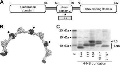
Fig. 4.
5.5T7 protein binds the central H-NS dimerization domain contained within residues 60 to 80. (A) Diagram of the H-NS molecule showing the approximate boundaries of each distinct domain within H-NS. (B) Structure of H-NS helical multimer (8 H-NS monomers) as recently solved by Arold et al. (2). The central dimerization domains that interact with gp5.5T7 are shown in dark gray and indicated with a star. (C) Coomassie-stained SDS-PAGE of the interaction between gp5.5T7 and various H-NS6His truncations after coexpression and purification over nickel resin. The positions of gp5.5T7 and various H-NS truncations are indicated.

