
Figure 2.

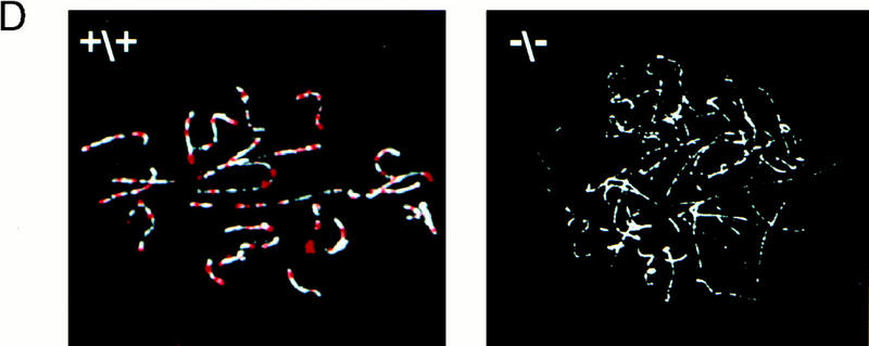
Generation of Msh4 mutant mice. (A) Gene-targeting strategy. Schematic representation of the Msh4 wild-type gene locus, the pMsh4Ex4 targeting construct and the targeted Msh4 locus. The exons are shown as black boxes. The PCR primers located in the PGK hygromycin cassette and in exon 6 that were used for detecting the gene-targeting events are indicated by arrows connected by a dotted line. The diagnostic BglII digestion products for the wild-type Msh4 locus and the modified Msh4 locus that are recognized by the hybridization probe are shown. (B) Southern blot hybridization of DNA from mice of the F2 generation. Tail DNA was digested with BglII and hybridized with the probe shown in A. The 5.2-kb band corresponds to the wild type and the 3.6-kb band corresponds to the targeted allele. (+/+) Wild type; (+/−) heterozygous; (−/−) homozygous. (C) Detection of Msh4 by Northern blot analysis. Testis poly(A) RNA of the different genotypes was analyzed with a cDNA probe corresponding to the entire Msh4-coding sequence. A human β-actin-specific probe was used as a control. (+/+) Wild type; (+/−) heterozygous; (−/−) homozygous. (D) Detection of MSH4 protein on meiotic chromosomes. Immunofluorescent colocalization of MSH4 (red) and the synaptonemal complex protein COR1 (white) on chromosome spreads of spermatocytes from 20-day-old males. (+/+) Chromosome spread from wild-type spermatocytes; (−/−) chromosome spread from homozygous mutant spermatocytes.