
Figure 3.
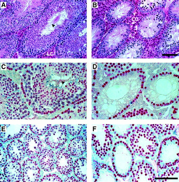
Testis morphology in Msh4+/+ and Msh4−/− males. (A,C,E) Wild-type males. (B,D,F) Msh4−/− mutant males. Hematoxylin and eosin staining (A,B) and immunohistochemical localization of GCNA1-positive spermatogonia and spermatocytes (C,D) in sections of adult testis. (E,F) GCNA1 localization of spermatogonia and spermatocytes at the end of the first wave of prophase I at day 23 pp. (LC) Leydig cells. Arrowheads show mature spermatozoa within the lumen of testes from wild-type adult males. (A–F) Bar, 200 μm.