Fig. 5.
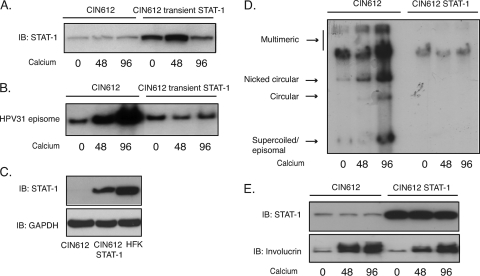
Restoration of STAT-1 levels blocks HPV genome amplification upon keratinocyte differentiation. (A) Western blot analysis of STAT-1 protein levels in CIN612 cells or in cells transiently expressing STAT-1 following differentiation in high-calcium medium for the indicated times. (B) Southern blot analysis for HPV31 episomes in CIN612 cells or the cells transiently expressing STAT-1 following differentiation in high-calcium medium for indicated times (h). (C) Western blot analysis for STAT-1 and involucrin protein levels in CIN612 cells or CIN612 cells stably expressing STAT-1 following differentiation in calcium medium for the indicated times (h). (D) Southern blot analysis for HPV31 genome in CIN612 cells or the cells stably expressing STAT-1 following differentiation in high-calcium medium for the indicated times (h). (E) Western blot assay for STAT-1 expression in CIN612 cells, CIN612 cells stably expressing STAT-1, and HFK cells. All results are representative of observations from two or more independent experiments.

