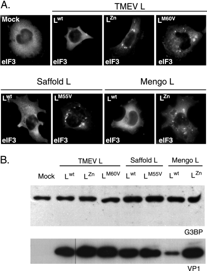
Fig. 7.
Inhibition of SG assembly is a common activity of cardiovirus L protein. (A) U3A cells were infected in parallel with a wild-type TMEV or with TMEV recombinants expressing TMEV LZn, TMEV LM60V, SAFV-2 Lwt, SAFV-2 LM55V, mengovirus Lwt, or mengovirus LZn. At 8 hours postinfection cells were fixed and processed for coimmunolabeling of VP1 (not shown) and eIF3. Each picture shows the distribution of eIF3 in a VP1-positive cell (not shown), except for the control, which was VP1 negative. (B) Western blot detection of G3BP in extracts from U3A cells infected as in panel A. No sign of G3BP cleavage or decrease in band intensity was observed when comparing wild-type and L-mutant viruses. Note that lower VP1 expression in SPA24 (Lwt mengovirus)-infected cells was noticed previously (23) and is thought to result from an altered synchronism between viral replication and L activity, which can indirectly impair viral replication.

