
Fig. 3.
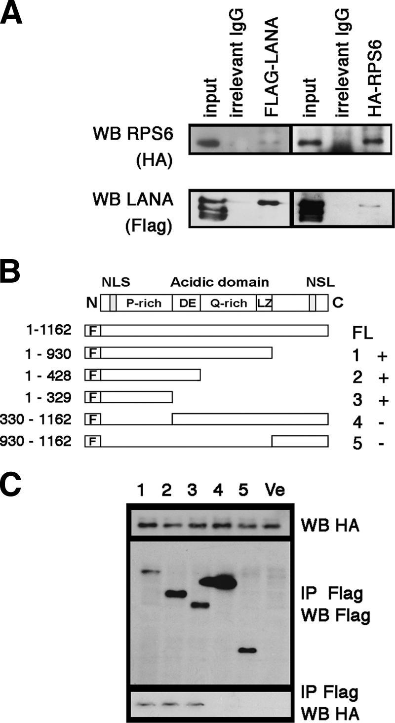
Analysis of the interaction of LANA and RPS6 in HeLa cells after transfection. (A) Coimmunoprecipitation of full-length FLAG-tagged LANA and HA-tagged S6 after cotransfection into HeLa cells. After cotransfection, protein extract was immunoprecipitated with the indicated reagents. The lanes represent input, IgG only, anti-FLAG, and anti-HA (HA). Anti-FLAG antibodies and anti-HA antibodies were used for Western blot (WB) analysis, as indicated on the left. (B) Schematic diagram of mutant LANA proteins used. All segments were fused with FLAG tag at the N terminus of LANA. The numbers on the right indicate the lane numbers in panel C and the “+” or “−” symbols summarize whether or not an interaction was detected. (C) Coimmunoprecipitation assay of HA-RPS6 and different mutants of FLAG-tagged LANA. Anti-FLAG and anti-HA antibodies were used for immunoprecipitation (IP) or Western blotting (WB) as indicated. Ve, vector.