Fig. 4.
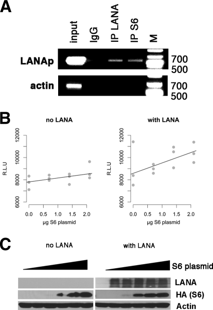
Complex of LANA and S6 binds to LANA promoter. (A) BC-3 cells were cross-linked and sonicated, and chromatin immunoprecipitation (IP) was performed with mouse anti-LANA (LN53) and mouse anti-RPS6 (54D2) monoclonal antibodies. IgG was used as a control. Inputs were 5% of lysates of each sample. The DNA fragments immunoprecipitated were amplified with the respective primers for the LANA promoter (LANAp) and β-actin. (B) Cotransfection of LANA and RPS6 into SLK cells increases the activity of a LANA promoter-luciferase reporter. Relative light units (RLU) are shown on the y axis, and the amount of S6 plasmid per transfection mixture is shown on the horizontal axis. All measurements were conducted in triplicate, and each is shown. The background (pGL3 vector) activity was below 100 RLU. Also shown are the regression lines (black), which demonstrate a linear dose dependence of LANA promoter activity on RPS6. (C) Western blot analysis of SLK cells cotransfected with increasing amounts of HA-tagged RPS6 expression vector in the absence or presence of LANA. A Western blot for actin is shown as a loading control.

